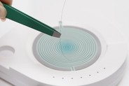
Hair bulb follicle through the process of the S-Drive.
The S-Drive is a peripheral computer device, designed to digitize the epigenetic data found in the hair bulb follicle through the process of the S-Drive.The gathered hair folical information (excluding the clients name and address) is then encrypted by the S Drive program, before being securely sent through an internet connection, to an encrypted server at our informational center located in Hamburg, Germany. Here the data is decoded, translated and scrutinized through an array of logarithms connected to an Ai processor, which then generates the completed commentary data pack. This personalized commentary data pack is returned securely through a separate server, linked to the practitioner’s coded S-Drive, within 15 minutes. The practitioner’s S-Drive program then automatically unpacks the data and creates the personalized PDF report. The S-Drive program then adds the client’s name and the practitioner’s details to the PDF report.
The multi-page report can be viewed, printed or forwarded as an email attachment to the client. The wide ranging observational reports provides the practitioner with additional underlying observations, to assist in decision making and determining supplementary protocols, in order to optimize the state of wellness and wellbeing of the client. The technology utilizes the principles of Homeostasis and epigenetics. The information is deduced by using four hair bulbs / follicles, plucked from the occipital (rear) area of the skull. The resulting personalized nutritional reports includes information on 12 Key Nutritional Indicators, plus additional pages on Gut, Immune and Circulatory support indicators, as well as Resistance and Environmental indicators. The 30 plus page report also highlights Food & Additive restrictions. All of the indicators are listed in an easy to read comprehensible report.

A 90 day optimized food plan is also part of the report which clients have found very easy to follow.
The technology was developed in order to optimize the wellbeing of the human body, as many of us are living in a sub-optimal functional state and more than often, well below our genetic and chronological potential. Intrinsically, poor functional genomic expressions are reflections of the life styles and environmental factors that we are exposed to.
The environment, our nutritional food intake, mental and emotional states play a major role in gene expression. Consequently, epigenetic influencers play a major role in our wellbeing.
The S-Drive process has been safety assessed and certified to electrical standards CE-(EU)ROHS Directive 2011/65(EU), FCC (USA)and ETL/UL(USA) and Canada, equal to those used for laboratory equipment, and does not emit any frequencies. Importantly, the S-Drive is not intended to diagnose, treat, cure, or prevent disease, as expressly stated on the reports that are generated from the S-Drive. The S-Drive is fully compliant with FDA guidance 1300013 (UCM429674).
The S Drive is operated in 67 countries by: wellbeing practitioners, sports professionals, fitness and beauty centres, nutritional professionals, Trichology Centres, Dietary Advisors, Weight Loss Centres, Spa Resorts and preventative organisations.
EPIGENETICS OBSERVATIONS
In order to grasp the significance of the technology, it is important to refer to the terms known as Homeostasis and Epigenetics. Epigeneticsis the study of changes in organisms caused by modification of the gene expression, rather than the alteration of the genetic code itself. Epigenetics has transformed the way we think about the consequences of genome hereditary. Homeostasis is the ability to maintain a relatively stable internal state that persists, despite changes in the world outside. All living organisms, from plants to puppies to people, must regulate their internal environment to process energy and ultimately survive. Most of us require ‘empowering nutritional food information’ in order to claim back the control over our bodies and our daily performance. Most humans do not understand that environmental and nutritional factors can directly impact our performances and wellbeing. Providing this information to a client can assist the practitioner in optimizing aspects of the client’s life. For the clinician, it is also important to discover the environmental impacts on the client.
Traditionally, hair shaft analysis has been utilized in forensics and environmental toxicology, as a tool to detect heavy metal toxicity and to trace certain contaminants that build over time, which may no longer be present in body fluids such as blood and urine. The forensic technique of hair analysis has been assessing a grid of different characteristics of hair, by comparative analysis between a known and unknown hair sample in order to establish a link. For example it compares hair found at the scene of a crime with samples taken from the suspect. Electron microscope were used until recentley, for forensic hair analysis. However light field microscopy is more often used to determine the color and to differ between animal and human hair. DNA analysis is also used and mass spectrum analyses (spectroscopy) to determine other elements contained.


The S-Drive technology however, relies on hair & bulb resonance information, combined with remote computer calculations, in order to reveal challenges that the underlying systems of the body have been experiencing over time. The technology provides a personalized, real-time overview of the underlying metabolic conditions, which determine wellness and wellbeing. This is achieved by digitizing the information found in four strands of hair and hair bulbs. The hair and bulb follicles have proven to be effective biomarkers, as they accumulate information from Homeostasis and epigenetic over time. This reflects a variety of underlying metabolic processes, often before the expression of a symptom.
The Homeostasis and epigenetic information can be reflected and analyzed using scanners, computer-based evaluation programming and Ai technology. The speed of computing technology, combined with remote servers, provides an effective operating platform. Emotions play a major role on Homeostasis and Epigenetics, since emotions are energies which are constantly moving and thus changing. The word emotion is an acronym that means ‘energy in motion’ that is energy that needs to move. We express emotion as feelings but feelings are not emotions, they are a way of expressing our state of being at any given time. Emotion is the energy that drives our thoughts so that they can materialize and thus express themselves, as well as a simple thought triggers an emotion capable of changing our entire physiology. Therefore, Homeostasis and Epigenetic influencers are a non-linear informational systems.
Strands of hair with their bulbs contain a wide range of historic Homeostasis and Epigenetic information concerning the metabolic parameters such as: amino acid demands, vitamins and mineral stresses, omega fatty acids requirements, sensitivities due to certain foods or the exposure to electromagnetic frequency (EMF) and extremely low electromagnetic frequencies (ELF), or ttoxic loads from the environment. These Homeostasis and Epigenetic markers can be used to highlight disharmony within the human body. Cellular and energetic stressors. *Note, disharmony may result in the establishment of chronic degeneration, caused by microbiological opportunism due to bacterial, fungal and parasitic infestations and environmental toxins, including heavy metal toxicity.
OVERVIEW OF THE OBSERVATIONAL INFORMATION
The human body is composed of trillions of cells. Each cell has its own genome represented by DNA and genes, which are packed in 44 autosomes and 2 sexual chromosomes. The human genome is made of 25,000 genes and over 150,000 proteins.
The Genome does not answer every question of wellbeing as it is thought that 98% of gene expression is dictated by the epigenome. This suggests that only 2% of the human genomic expression is under the influence of the genotype. Coding Genes make up 2 to 3% of the entire DNA, the rest is referred to as non-coding DNA, and that accounts for up to 98% of our physical self. The observational value of Homeostasis and Epigenetic information is therefore important.
Homeostasis and Epigenetic information includes the experiences of our ancestors, our current diets and lifestyles, as well as our thoughts and beliefs. Beliefs and thoughts have a direct effect in our emotions, mediated by the synthesis of neuropeptides from the enteric nervous system. Thus they exhibit a direct impact on our physiology such as the so called placebo effect (epigenetics in action). There is also the nocebo effect.
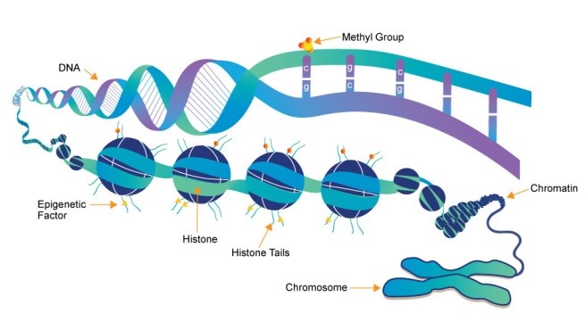
Homeostasis and Epigenetic observations include the study of gene expression under the influence of informational signals emanating from the micro and the macro environment. In this modeling, the phenotype changes but the genotype remains the same, due to methylations of the histones. Homodynamic and epigenetic mechanisms are affected by several factors and processes including development in utero and in childhood, environmental chemicals, drugs and pharmaceuticals, aging and diet. DNA methylation is what occurs when methyl groups, and epigenetic factors found in some dietary sources, can tag DNA and activate or repress genes.
Another way to look at Genetics is that the genes read the environmental factors via messenger ribonucleic acid (mRNA), resulting in the synthesis of proteins which are directly related to the epigenome (Doerfler Walter and Petra Böhm Springer eds. 145-175. ISBN: 978-3-319-27186-6. 2016). The proteins responsible for epigenetics are histones and chromatin, from which chromosomes and autosomes are formed. The non-coded DNA is what is commonly referred to as “Junk DNA”, and its amount varies from specie to specie. In the human being there is up to 98% non-coding DNA or epigenetics. (Carey N (2015) Junk DNA: A Journey Through the Dark Matter of the Genome. Icon Books Ltd. Chapter 3. ISBN: 978-184831-826-7)
Humans can no longer be considered as mechanical machines, controlled by genetic inheritance, the genotype. Observational Homeostasis and epigenetic information provides information to allow each human to take control of their own wellbeing. We now understand how to influence our ‘epigenomic environment’, in order to improve the impact of physical, mental and emotional issues.
Homeostasis and epigenetic informational knowledge empowers clinicians and clients.
The observations provide instant access to nutritional knowledge and key nutritional factors, which can be used to bring about optimization, using diet nutritional and lifestyle changes to bring about optimal gene expression. The changes are intended to stimulate positive and relevant gene expression. The life style suggestions can impact the system to promote physiological efficiency and metabolic efficacy.
INTRODUCTION TO HAIR & BULBS AS BIO MARKERS
Hair shafts and their bulbs/follicles store former metabolic and nutritional data, which reveals sensory bio-information.
Hair can be found on all of the major visible surfaces of the body. It is also the only body structure that is completely renewable without scarring.
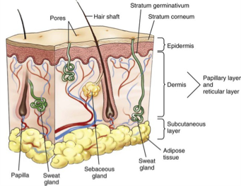
The ectoderm is the extreme outer layer of the neural tube, which forms at the end of the 4th week of gestation. This develops the hair as well as the rest of integumentary system, which comprises the skin, the nails, the teeth along with the nervous system comprising the brain, the spinal cord and nerves. A developing fetus has all of its hair follicles created by the 22nd week of pregnancy. At this time, there are at least 5 million follicles over the body; one million of these are on the head, this will be the largest number of follicles we will ever have, as follicles cannot be added later in life. When we grow older, the size of the body increases and the density of hair follicles on the skin decreases.
HAIR SHAFT and BULB ANATOMY
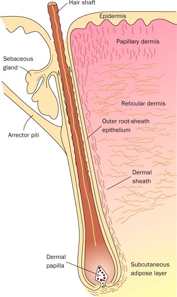
Hair has two separate structures - the follicle within the skin and the shaft which grows from it that we see. As shown in figures 1 and 2. At the base of the follicle is the so-called dermal papilla. It contains capillaries (tiny blood vessels), that feed the structure.
The living part of the hair is the follicle and is the only part fed directly by the capillaries. The cells in the bulb divide every 23 to 72 hours, faster than any other cells in the body. Due to this, the hair can be used to trace metabolic deficiencies, a possible storage of heavy metals, metabolic toxicity, etc. This array of information can be used to trace the individual metabolic history.
The follicle is surrounded by two sheaths - an inner and outer sheath. These sheaths protect and mold the growing hair shaft. The inner sheath follows the hair shaft and ends below the opening of a sebaceous (oil) gland, and sometimes an apocrine (scent) gland as shown in figure 2. The outer sheath continues all the way up to the gland. A muscle called the erector pili muscle attaches below the gland to a fibrous layer around the outer sheath. When this muscle contracts, it causes the hair to stand up (goose bumps).
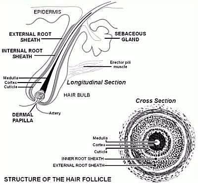
The sebaceous gland is important, since it produces sebum as a natural conditioner. More sebum is produced after puberty. The hair shaft is composed of hard protein called keratin, without actual living cells. The production of this tissue decreases in men, but not as much in women. The hair’s structure is divided into three layers: the inner layer is called “medulla” and may not be present. The next layer is the “cortex“ and the outer layer is the “cuticle“. The cortex layer makes up the majority of the hair shaft. The cuticle is formed by tightly packed scales in an overlapping structure, similar to roof shingles. Most hair conditioning products attempt to affect the cuticle.
There is a pigment called melanin, that is distributed throughout the cortex and medulla giving the hair its characteristic color. The more melanin, the darker the hair’s color appears. The hair is a non-living integumentary structure. It has an ectodermal origin the same as the skin, the connective tissue and the nervous tissue, that includes the brain, spine cranial and spinal nerves. The hair and bulbs have a sensory structure that is connected to the erector pili muscle, the smallest muscles in the body, which contraction allows the hair to rise. (Embryology of the Integumentary system (2016) https://embryology.med.unsw.edu.au/embryology/index.php/Integumentary_System_Development)
The hair follicle is in contact with the dermis and it has living cells such as melanocytes, keratinocytes and fibroblasts. All these cells contain DNA in their nucleus. The hair is made of a structural protein called keratin and its color is due to a protein called melanin. (Yasuyuki Amoh, Lingna Li, Kensei Katsuoka, Robert M Hoffman Embryonic development of hair follicle pluripotent stem (hfPS) cells. Med Mol Morphol: 2010, 43(2);123-7)
The hair stores information emanating from the micro and macro environment, (https://lsda.jsc.nasa.gov/Experiment/exper/1517 ) and also can store toxins such as heavy metals, that if set free could harm the body. The hair is regarded as a structure providing protection, acting as a waste bin, storing substances such as byproducts of cellular metabolism that could potentially harm the body, as well as protecting us from the harmful effect of chemicals that are applied to the organism by means of cosmetics. (M. I. Szynkowska, A. Pawlaczyk, E. Wojciechowska, S. Sypniewski, T. Paryjczak (2009).
Human Hair as a Biomarker in Assessing Exposure to Toxic Metals.Polish J. of Environ. Stud. Vol. 18, No. 6, 1151-1161 ) The hair follicle contains enzymes known as aromatases and 19-hydroxylases that change male hormones into female hormones. That is Androgens such as Testosterone into Estrogen such as Estradiol. So, the hair plays a role in neuroendocrine regulation. The hair takes up to 80 days to surface, and go beyond the hair shaft.
REFERENCES
- Gaillard, Y., Pepin, G. Testing Hair for pharmaceuticals, J. Chromatogr. B 733 (1999) 231–246.
- Orozco, C., Hair Pathology and Hair Physiology. Lecture Notes (unpublished). Australian Institute of Applied Sciences. Brisbane Australia, 2010.
- Orozco C, Biochemistry and Nutrition. Lectures Notes (unpublished). Australian Institute of Applied Sciences. Brisbane Australia, 2011. •http://rogeriannursingscience.wikispaces.com/Chapter+5+T he+Scien ce+of+Unitary+Human+Beings+Principles. Downloaded on the 10 April 2014.
- Henderson, G.L., Harkey, M.R., Jones, R.T ., Analysis of hair for opioids” (eds. Edward. J. Cone, Ph.D., Michael. J. Welch, Ph.D., and M. Beth Grigson Babecki, M.A.), "Hair T esting for Drugs of Abuse: International Research on Standards and Technology", 1995, p. 91-120. NIH Publication No. 95-3727
- Kintz, P., Bioanalytical procedures for detection of chemical agents in hair in the case of drug-facilitated crimes. Anal Bioanal Chem. 388, 7 (2007) 1467-74.
- Nakahara, Y., Hair analysis for abused and therapeutic drugs, J. Chromatogr. B 733 (1999) 161–180.
- Romolo, Lopez, F .S., Rotolo, M.C., Palmi, I., Lopez, A., Optimized conditions for simultaneous determination of opiates, cocaine and benzoylecgonine in hair samples by GC-MS. Forensic Science International (2003), 138(1-3), 17-26.
- Sachs, H. Kintz, P., Testing for drugs in hair. Critical review of chromatographic procedures since 1992, J. Chromatogr. B 713 (1998) 147– 161.
- Pragst F., Balikova M.A.: State of the art in hair analysis for detection of drugs and alcohol abuse; Clinica Chimic Acta 370 2006 17-49.
- Pragst F., Auwärter V., Kiessling B., Dyes C.: Wipe-test and patch- test ror alcohol misuse based on the concentration ratio of fatty acid ethyl esters and squalen CFAEE/CSQ in skin surface lipids. Forensic Sci Int 2004; 143:77-86.
- Leklem JE. Vitamin B6. In: Shils ME, Olson JA, Shike M, Ross AC, ed. Modern Nutrition in Health and Disease. 9th ed. Baltimore: Williams and Wilkins, 1999: 413-421.
- Bender DA. Vitamin B6 requirements and recommendations. Eur J Clin Nutr 1989 ;43:289-309.
- Gerster H. The importance of vitamin B6 for development of the infant. Human medical and animal experiment studies. Z Ernahrungswiss 1996; 35:309-17.
- Bender DA. Novel functions of vitamin B6. Proc Nutr Soc 1994; 53:625-30.
- Chandra R and Sudhakaran L. Regulation of immune responses by Vitamin B6. NY Acad Sci 1990; 585:404-423.
- Trakatellis A, Dimitriadou A, Trakatelli M. Pyridoxine deficiency: New approaches in immunosuppression and chemotherapy. Postgrad Med J1997;73:617-22.
- Shibata K, Mushiage M, Kondo T, Hayakawa T, Tsuge H. Effects of vitamin B6 deficiency on the conversion ratio of tryptophan to niacin. Biosci Biotechnol Biochem 1995; 59:2060-3.
- Leyland DM and Beynon RJ. The expression of glycogen phosphorylase in normal and dystrophic muscle. Biochem J 1991; 278:113-7.
- Oka T, Komori N, Kuwahata M, Suzuki I, Okada M, Natori Y. Effect of vitamin B6 deficiency on the expression of glycogen phosphorylase mRNA in rat liver and skeletal muscle. Experientia 1994; 50:127-9.
- Okada M, Ishikawa K, Watanabe K. Effect of vitamin B6 deficiency on glycogen metabolism in the skeletal muscle, heart, and liver of rats. J Nutr Sci Vitaminol (Tokyo) 1991; 37:349-57.
- DeLuca HF and Zierold C. Mechanisms and functions of vitamin D. Nutr Rev 1998;56:S4-10.
- Reichel H, Koeffler H, Norman AW. The role of vitamin D endocrine system in health and disease. N Engl J Med 1989;320:980-91.
- Van den Berg H. Bioavailability of vitamin D. Eur J Clin Nutr 1997;51 Suppl 1:S76-9.
- Institute of Medicine, Food and Nutrition Board. Dietary Reference Intakes: Calcium, Phosphorus, Magnesium, Vitamin D and Fluoride. National Academy Press, Washington, DC, 1999.
- Goldring SR, Krane S, Avioli LV. Disorders of calcification: Osteomalacia and rickets. In: LJ D, ed. Endocrinology. 3rd ed. Philadelphia: W B Saunders, 1995:1204-27.
- Favus MJ and Christakos S. Primer on the Metabolic Bone Diseases and Disorders of Mineral Metabolism. 3rd ed. Philadelphia, PA: Lippincott- Raven, 1996.
- Holick MF. Evolution and function of vitamin D. Recent Results. Cancer Res 2003;164:3-28.
- Biser-Rohrbaugh A, Hadley-Miller N. Vitamin D deficiency in breast-fed toddlers. J of Pediatr Orthaped 2001;21:508-11.
- Wharton B and Bishop N. Rickets. The Lancet 2003;362:1389-1400.
- Osiecki H, The Nutrient Bible. 2004; 31-33.
- Shils M, Olson JA, Shike M, Ross AC, eds. Modern Nutrition in Health and Disease. 9th ed. Baltimore: Williams & Wilkins; 1999:401–411.
- Jacob R, Swenseid M. Niacin. In: Ziegler EE, Filer LJ, eds. Present Knowledge in Nutrition. 7th ed. Washington D.C: ILSI Press; 1996:185-190.
- Jacobson MK, Jacobson EL. Discovering new ADP-ribose polymer cycles: protecting the genome and more. Trends Biochem Sci. 1999;24(11):415-417.
- Lander ES (2011). "Initial impact of the sequencing of the human genome". Nature 479 (7333): 187–197.
- Cheung P, Lau P (2005) Epigenetic regulation by histone methylation and histone variants. Mol Endocrinol Mar;19(3):563-73.

Locate Our Optimize Report Provider
Find a providerBecome Provider
Be a providerAll Cell Wellbeing reports are provided for educational and informational purposes only. They are non-diagnostic, do not provide medical or veterinary advice, and are not intended to diagnose, treat, cure, or prevent any disease or condition.
None of these statements have been evaluated by the FDA. This product is not intended to diagnose, treat, cure or prevent any disease or condition. It is intended to provide nutritional food information. The digital process uses We use In-Vivo rather than In-Vitro technology and therefore does not provide reproducible indicators as it reflects the constant changing epigenetic environment at the quantum biological level. For this reason, nutritional food optimization should only be considered every 90 days. it is NOT recommended that a new Optimized report be created within this period.